

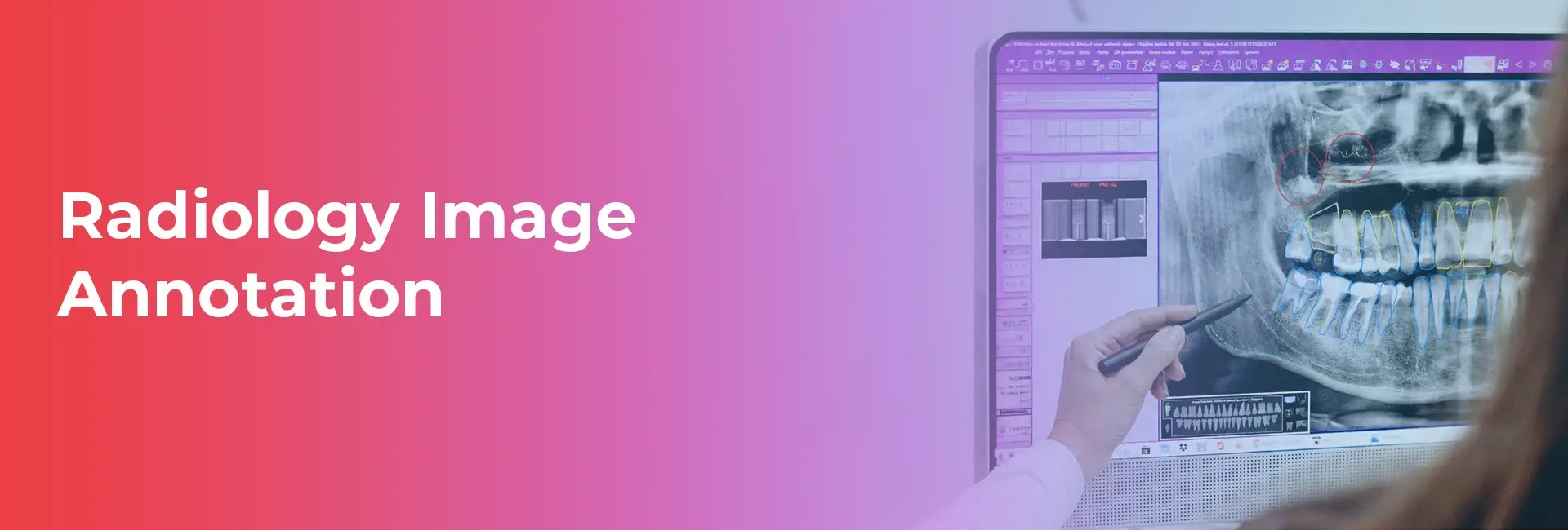
- What Is Radiology Image Annotation?
- Why Radiology Image Annotation Matters for Healthcare AI
- Common Types of Radiology Image Annotation
- Key Challenges in Radiology Image Annotation
- Best Practices for High-Quality Radiology Image Annotation
- How Radiology Image Annotation Powers Modern AI Applications
- Quality Assurance in Radiology Image Annotation
- Choosing the Right Partner
- The Future of Radiology Image Annotation
- Moving Forward with Healthcare AI Data
- FAQs
Radiology Image Annotation: Building Accurate Medical AI
The adoption of artificial intelligence in medical imaging and diagnostics is accelerating rapidly. Healthcare organizations and AI startups are developing powerful tools to detect diseases earlier, improve patient outcomes, and streamline clinical workflows. However, the performance of these machine learning models relies entirely on the quality of their training data. High-quality medical imaging data is critical. Without it, even the most advanced algorithms cannot function safely or effectively in a clinical setting.
Radiology image annotation serves as the vital bridge between raw medical data and intelligent diagnostic systems. By carefully labeling X-rays, MRIs, and CT scans, medical experts teach AI models to recognize complex anatomical structures and subtle disease markers. Accurate annotations directly improve disease detection, support precise medical image segmentation, and ultimately enhance clinical decision-making. Developing these models requires a specialized approach, which is why organizations turn to Macgence for expert-led radiology image annotation services tailored to healthcare AI development.
What Is Radiology Image Annotation?
Radiology image annotation is the process of labeling and classifying medical images to train artificial intelligence and machine learning models. Unlike general image annotation, which might involve identifying everyday objects like cars or trees, medical image annotation requires a deep understanding of human anatomy and pathology.
During this process, experts mark specific regions of interest within a scan. They might highlight a tumor, trace the boundaries of an organ, or classify a bone fracture. This expert-guided annotation is crucial for creating reliable healthcare datasets. An AI model trained on poorly labeled medical images could produce life-threatening diagnostic errors.
Annotators work across a wide variety of common imaging modalities, including:
- X-rays
- CT Scans
- MRI Scans
- Ultrasound Images
- PET Scans
- Mammography Images
Why Radiology Image Annotation Matters for Healthcare AI
Radiology image annotation matters because it directly enables AI-powered disease diagnosis. When algorithms learn from accurately labeled data, they can support the early detection of abnormalities that might be invisible to the naked eye. This process improves medical image segmentation and classification, leading to enhanced model accuracy and reliability. Ultimately, these AI tools reduce the diagnostic workload for healthcare professionals, allowing radiologists to focus on complex cases and patient care.
Some common example applications include:
- Tumor detection
- Lung disease diagnosis
- Brain lesion segmentation
- Fracture detection
- Breast cancer screening
Common Types of Radiology Image Annotation
Bounding Box Annotation
Bounding box annotation involves drawing a rectangular box around a specific abnormality, such as a lung nodule or a fractured bone. It helps identify and localize abnormalities quickly and is highly suitable for training object detection models.
Semantic Segmentation
Semantic segmentation assigns a specific class to every single pixel in an image. This pixel-level labeling of organs, tissues, and lesions is heavily used in precision diagnostics, where understanding the exact boundaries of a disease region is necessary.
Instance Segmentation
Instance segmentation goes a step further by differentiating multiple objects within a single image. If a scan shows multiple distinct tumors, this technique identifies each one individually. It is particularly useful for tumor counting and precise organ identification.
Landmark Annotation
Landmark annotation focuses on marking specific anatomical points or structures across a medical scan. Clinicians and researchers use this technique for surgical planning, tracking anatomical changes over time, and taking exact medical measurements.
Polygon Annotation
Polygon annotation provides precise outlining of highly irregular structures. Since biological anomalies rarely form perfect squares, polygons allow annotators to trace the exact shape of complex disease regions, ensuring the AI model understands the true footprint of the pathology.
Key Challenges in Radiology Image Annotation
Requirement for Medical Expertise
The most significant hurdle is the need for radiologists and trained medical annotators. Medical images are highly technical, and the high cost of expert involvement can strain project budgets.
Complex Medical Imaging Data
Human anatomy and disease presentations feature enormous variability. Multi-dimensional imaging complexity, such as 3D MRI or CT volumes, adds a heavy layer of difficulty compared to standard 2D image labeling.
Annotation Consistency
Achieving high agreement among multiple annotators, known as inter-observer variability, is difficult. Maintaining consistent quality across large datasets requires rigorous oversight and continuous training.
Data Privacy and Compliance
Medical images contain sensitive patient health information. Teams must navigate HIPAA regulations, GDPR, and other healthcare data frameworks to ensure secure annotation workflows and protect patient privacy.
Scalability Issues
There is a growing demand for massively large, annotated medical datasets to train sophisticated foundation models. Balancing the speed of data processing with the strict accuracy required for clinical use remains a major industry challenge.
Best Practices for High-Quality Radiology Image Annotation
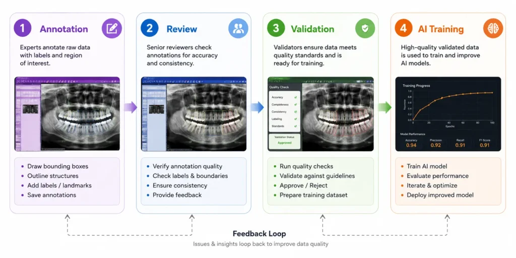
To overcome these challenges and produce reliable training data, AI teams must adhere to a strict set of best practices. First, it is essential to develop detailed annotation guidelines before starting any project. Teams should use certified medical experts for data validation to ensure clinical accuracy.
Organizations must implement multi-layer quality assurance protocols and conduct consensus reviews for highly complex cases. Maintaining standardized labeling protocols guarantees that the data remains uniform. Furthermore, teams should utilize specialized annotation tools optimized for DICOM formats and medical imaging workflows.
Pro Tip: Incorporate continuous feedback loops between your AI engineers and medical annotators. Reviewing model predictions against human annotations helps identify recurring errors and improves annotation quality over time.
How Radiology Image Annotation Powers Modern AI Applications
Computer-Aided Diagnosis (CAD)
Annotated data trains CAD systems to flag potential issues on a scan, providing automated detection assistance to doctors reviewing hundreds of images daily.
Disease Classification Models
By feeding labeled examples of different pathologies into an algorithm, developers create models capable of identifying disease severity and specific types of conditions accurately.
Organ Segmentation
Detailed segmentation models help map out internal anatomy automatically, which is vital for radiation treatment planning and robotic surgical assistance.
Predictive Healthcare Analytics
Radiology image annotation contributes to predictive models that track disease progression, ultimately supporting customized, personalized medicine for patients.
Clinical Research and Drug Development
Large annotated datasets allow researchers to analyze how experimental treatments affect tumors and tissues, accelerating medical discoveries and pharmaceutical testing.
Quality Assurance in Radiology Image Annotation
Building a medical AI model without rigorous quality control is incredibly risky. Effective quality assurance requires multi-stage review workflows where every annotation passes through several checks. A core component of this is the radiologist verification process, ensuring the labels make clinical sense.
Teams also use automated quality checks to catch simple formatting errors or missing labels. Regular annotation consistency audits help keep large teams aligned. Finally, comprehensive dataset validation before model training ensures the AI algorithm learns from the best possible information.
Choosing the Right Partner
Selecting the right partner can make or break your healthcare AI project. Key factors to consider include the vendor’s access to true medical domain experts and their proven experience with healthcare AI projects. Data security and compliance standards must be flawless. You should also evaluate their scalability for large imaging datasets, their internal quality assurance framework, and their support for multiple annotation formats and medical modalities.
The Macgence Advantage
Macgence provides expert-led annotation teams capable of handling the most complex medical imaging workflows. By implementing rigorous quality control processes, Macgence delivers highly accurate, secure, and scalable healthcare AI data solutions customized to fit the exact needs of your machine learning models.
The Future of Radiology Image Annotation
The field of medical data labeling is evolving quickly. Emerging trends include the rise of AI-assisted annotation tools that pre-label images, leaving human experts to refine and verify the results through human-in-the-loop workflows. There is also a major push toward advanced 3D medical image annotation and the development of foundation models specifically built for medical imaging. These innovations will lead to much faster dataset creation for healthcare AI innovation.
The industry outlook is exceptionally strong. As clinical environments continue increasing their integration of AI into daily workflows, the global demand for high-quality, expertly annotated radiology datasets will only continue to grow.
Moving Forward with Healthcare AI Data
Radiology image annotation is the absolute foundation of developing safe, reliable healthcare AI systems. There is a direct and unbreakable connection between the quality of your annotation data and your model’s real-world clinical performance. As the industry advances, the need for expert-driven, strictly compliant, and highly scalable annotation workflows has never been more urgent. By partnering with experienced professionals, you secure the data integrity necessary to change the landscape of modern medicine. Macgence stands ready as a trusted partner, offering the expertise and secure workflows required for elite medical image annotation and comprehensive healthcare AI data solutions.
FAQs
Ans: – It is the process of labeling and highlighting specific anatomical structures or abnormalities in medical images to train artificial intelligence models.
Ans: – Accurate annotations teach AI algorithms how to identify diseases, map organs, and detect subtle abnormalities safely and effectively.
Ans: – Common modalities include X-rays, MRI scans, CT scans, ultrasound imaging, PET scans, and mammography.
Ans: – Because of the complex nature of the data, annotation is usually performed by trained medical professionals, certified annotators, and experienced radiologists.
Ans: – The most frequent techniques include bounding boxes, semantic segmentation, instance segmentation, polygon annotation, and landmark annotation.
Ans: – Major challenges include the high cost of medical experts, maintaining strict data privacy (HIPAA compliance), ensuring high inter-observer consistency, and scaling operations efficiently.
Ans: – Quality assurance involves multi-stage reviews and expert verification, ensuring that the labeled data is clinically accurate and completely reliable for model training.
Ans: – Macgence provides highly secure, scalable, and expert-led medical image annotation services, backed by rigorous quality control frameworks tailored to healthcare AI development.
 Previous Blog


You Might Like

June 8, 2026
Egocentric Video Annotation: Powering Embodied AI
The demand for embodied AI and robot learning is growing rapidly. Developers are shifting their focus from AI that simply observes the world to systems that actively interact with it. To achieve this, models need a different kind of training data. They need to see the world exactly as we do. Traditional third-person video datasets […]

June 5, 2026
Physical AI Datasets: The Foundation of Real-World Intelligent Systems
Traditional artificial intelligence systems have long operated entirely within the digital realm, processing text, generating images, and analyzing virtual data. However, a major shift is occurring as intelligent systems step out of the digital space and into the physical environment. This new era of Physical AI powers the machines that interact with our world—from self-driving […]

June 4, 2026
Building Global AI with Multilingual Audio Annotation Services
Voice-enabled artificial intelligence is rapidly transforming how businesses operate globally. From smart virtual assistants and voice search to advanced speech analytics and call center AI, speech technology is becoming a foundational element of customer interaction. To make these systems truly effective on a global scale, developers need accurate and diverse training data. High-quality multilingual audio […]